| FIG. 2 |
|
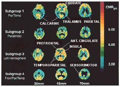
| PET scan images from four AD patients that best characterize four independent subgroups of metabolic patterns. Three planes are shown for each subject; left, at level of orbitofrontal cortex 30 mm above inferior orbitomeatal (IOM) line; middle, at level of basal ganglia 45 mm above IOM line; right, at level of centrum ovale 70 mm above IOM line. rCMRglc scale in mg/100 g/min. (From ref. 28.) |
| Back to Chapter |
published 2000