| Figure 3 |
|
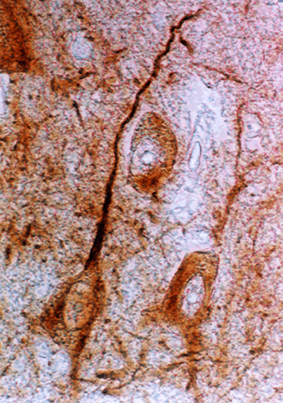
| NFM immunostaining of human entorhinal cortex neurons. Photomicrograph of stellate neurons from layer 2 of entorhinal cortex taken from post-mortem brain tissue of a diagnosed schizophrenic. Cells were stained with RMDO-20, a monoclonal antibody for mid-sized, poorly phosphorylated neurofilament, the staining of which is largely confined to somato-dendritic regions. Following immunohistochemical staining, individual stellate neurons were aspirated into patch electrodes for the in situ transcription of mRNAs followed by aRNA amplification. The expression of mRNAs from these cells was compared with the expression of mRNAs collected from neurons of age-matched controls using gene expression profiling and differential display. |
| Back to Chapter |
published 2000