| Figure 5 |
|
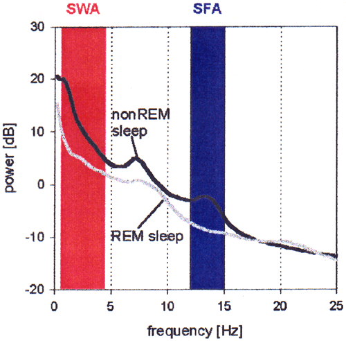
| Average EEG power spectra of nonREM (stages 1 to 4) and REM sleep (8 subjects, 1 night each). Power is plotted on a logarithmic scale (dB). Spectra were calculated for 4-s epochs (Hanning window) and five epochs were averaged and matched with the 20-s sleep scores. Only 20-s without artifacts were included. The frequency ranges of slow-wave activity (SWA, red area) and of spindle frequency activity (SFA, blue area) are marked. |
| Back to Chapter |
published 2000