| Figure 4 |
|
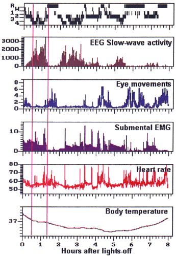
| Polygraphic recording of an 8-h nighttime sleep episode. From top to bottom: hypnogram with sleep scores (R: REM sleep; W: waking; 14: nonREM sleep stages 1 to 4); slow-wave activity (mV2; EEG power in the 0.75 4.5 Hz range); eye movements (ratio of mean RMS amplitudes in 20-s epochs of EOG and EEG); EMG (mV; mean RMS amplitude of 20-s epochs); heart rate (beats per minute); rectal temperature (tick marks 0.1° C). Pink vertical lines indicate the 20-s epochs depicted in Figure 2 and Figure 3. |
| Back to Chapter |
published 2000