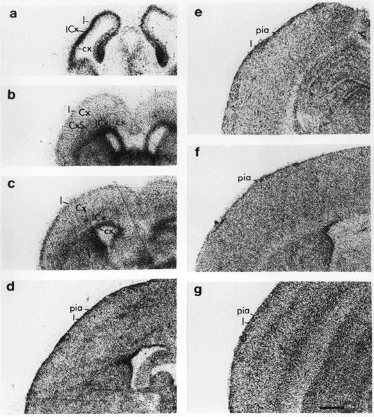
Figure 9. Autoradiographs of 125I-VIP binding in rat brain sections (in the coronal plane through the region of the cortex) during the development of the rat brain from E16 to 60 days of age. Autoradiography was performed as previously described (71). Bar = 2 mm. a: VIP binding in the E16 cortex showing dense binding in the intermediate layer of the developing cortex but none in the neuroepithelial layer. b: VIP binding in the E19 cortex illustrating the highest binding in the neuroepithelial layer and moderate levels of binding in layer 1 and the intermediate layer. c: VIP binding in the postnatal day (P) 0 cortex, showing that the neuroepithelial layer has narrowed but still has the highest binding, whereas binding is becoming more evenly distributed in other cortical layers. d: VIP binding in the P7 cortex, illustrating that, except for layer 1, moderately dense and evenly distributed VIP binding at this time obscures many details of regional anatomy. Very little neuroepithelium remains. e: VIP binding in the P14 cortex in which VIP binding is uniform except for increased binding in layer I. f: VIP binding in the P21 cortex showing homogenous density of binding. g: VIP binding in the 60-day-old adult rat brain illustrating the higher density of VIP binding in layers I, II, IV and VI of the cortex. Cx, cortical plate; cx, cortical neuroepithelium; CxS, cortical subplate; ICx, intermediate layer of the cortex. Cortical layers are denoted by Roman numerals. (Taken, with permission, from ref. 71)