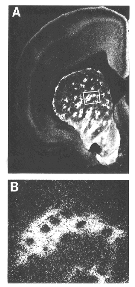
| Figure 3. |
|
|
| Dark-field photomicrograph illustrating distribution of mu opioid receptor binding sites in a coronal hemisection of rat forebrain labeled by [3H]-DAGO. Note the patchy appearance of binding within the striatum. B: Boxed area in A is shown at higher magnification. (Courtesy of Dr. Alfred Mansour.) |
published 2000